Tinea corporis is a skin disease due to infection of the skin by dermatophyte fungi. Dermatophytes are capable of colonizing keratinized tissue.
Three generas of dermatophytes are Trichophyton, Microsporum and Epidermophyton. Apart from the skin, dermatophytic infections can also affect the hair and nails.
The disease can be transmitted directly by contact with infected humans, animals or via fomites. Autoinoculation can also occur, a condition where an infection is transmitted from another part of the body in the same individual. Use of occlusive clothing, frequent skin-to-skin contact, hot and humid weather creates an environment which predispose to this condition.
Signs & Symptom
The patient would often complain of itchiness of the lesion and on inspection, the lesion is seen as an erythematous patch, slightly scaly with an active, serpiginous, erythematous margin. Small erythematous papules and pustules may be present at its periphery. If left untreated, the patch will enlarge and with the presence of its central clearing, it will appear ring-like or annular which is why it is often called ‘ringworm’.
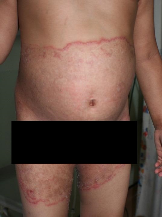 |
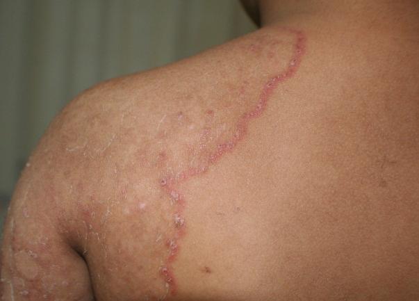 |
| The above photos showing lesions of tinea corporis: note the erythematous, slightly scaly macules with serpiginous erythematous margins and central clearing. | |
Tinea corporis can affect any parts of the body especially the body folds such as the armpits, thigh, buttocks, abdomen, etc.
The clinical diagnosis of tinea corporis can be confirmed by direct microscopy or fungal culture. In performing direct microscopic examination, the infected skin is scraped with the back of a disposable surgical blade, scrapings then smeared on a glass slide, mixed with drops of potassium hydroxide and observed under the microscope. Another technique is fungal culture, where the scrapings are sent to the lab, cultured in a special media and the species of proliferating fungal colony can be identified after a few days.
Complication
Identification of tinea corporis can be tricky in certain circumstances. Lesions that have been treated with steroid creams would have an altered appearance. This may lead to some difficulty in diagnosis and in this situation, it is called ‘tinea incognito’
Other skin diseases that may look similar to tinea corporis especially to the untrained eye include: psoriasis, discoid eczema, urticaria, Bowen’s disease, leprosy, secondary syphilis , etc. (see photos)
Skin conditions which may be confused as tinea corporis include:
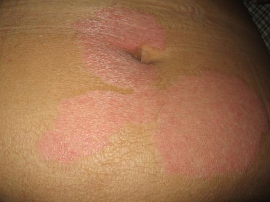 |
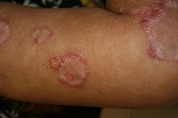 |
|
a) Psoriasis
|
|
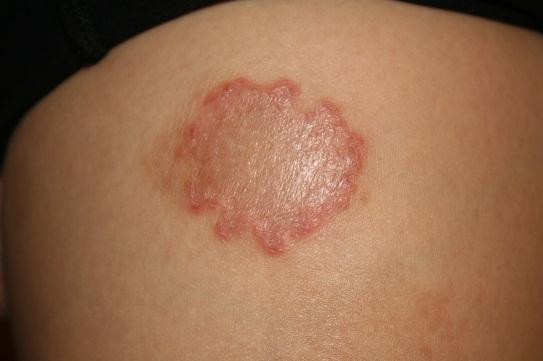 b) Discoid eczema |
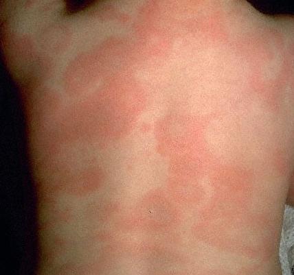 c) Urticaria |
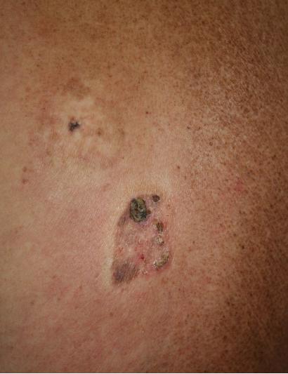 d) Bowen’s disease |
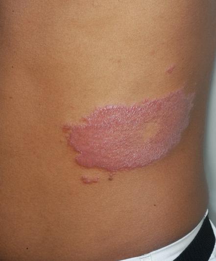 e) Leprosy |
Treatment
Tinea corporis can be cured with antifungal medications. The method of treatment depends on various factors, such as the size/extent of lesion, whether it is recurrent after initial treatment, not responsive to topical treatment, etc
If the lesion is limited to one or few areas, topical treatment with creams containing imidazole, allylamine, tolnaftate and cicloprox can be used. The correct application technique cannot be overemphasized. The cream has to be applied on the lesion and extended slightly beyond its active margin. Apply twice daily for two to four weeks, depending on the type of medication used.
If the tinea corporis is extensive, oral/systemic treatment would be a more practical choice. Oral antifungals that are available include, itraconazole, terbinafine and fluconazole. These medications have various side effects and may interact with other medications. Therefore, ensure that these medications are prescribed by medical practitioners who are familiar with its use.
Prevention
Prompt and adequate treatment is important to ensure remission and to prevent recurrence. Avoid sharing towels or personal clothing and ensure that the affected areas are kept dry.
Despite these steps, recurrences can still occur especially in individuals with predisposing factors such as poorly controlled diabetes mellitus or those who are immunosuppressed.
References
- Hay RJ, Ashbee HR.(2010). Burns T, Breatneach S, Cox N, Griffiths C. (Eds). Rook’s Textbook of Dermatology.(pp.1657- 1707). United Kingdom: Wiley-Blackwell
- Verma S, Heffernan MP. (2008)Wolff K, Goldsmith LA, Katz SI, Gilchrest BA, Paller AS, Leffell DJ. (Eds).Fitzpatrick’s Dermatology in General Medicine. (pp 1807-1821).USA: McGraw-Hill
| Last Reviewed | : | 23 August 2019 |
| Writer | : | Dr. Mazlin Baseri |
| Accreditor | : | Datin Dr. Asmah bt. Johar |
| Reviewer | : | Dr. Nazatul Shima bt. Abd Rahim |


