“There is a hole in front of my EAR?”
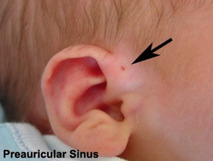 |
| Figure 1 : Preauricular Sinus (Source: https://s-media-cache ak0.pinimg.com/originals/e6/b1) |
Introduction
A preauricular sinus is a small opening or pit adjacent to the external ear. It is a congenital malformation that present at birth. Also known as a congenital auricular fistula, a congenital preauricular fistula, an ear pit or a preauricular cyst, usually becomes more apparent later in life.
Most people with preauricular sinus are asymptomatic and require no treatment. However, in a person with a repeated infection, excision of the sinus may be necessary.
The sinus may occur on both ears (bilateral) in 25-50% of cases, and they are more likely to be hereditary5. In one-sided (unilateral) cases, the left side is more commonly affected. Men and women are equally affected .
Clinical Features
Preauricular sinuses are frequently noted on routine physical examination as small pits or small openings adjacent to the external ear. It is usually located at the front part of the ear pinna. Other locations of preauricular sinus are rarer.
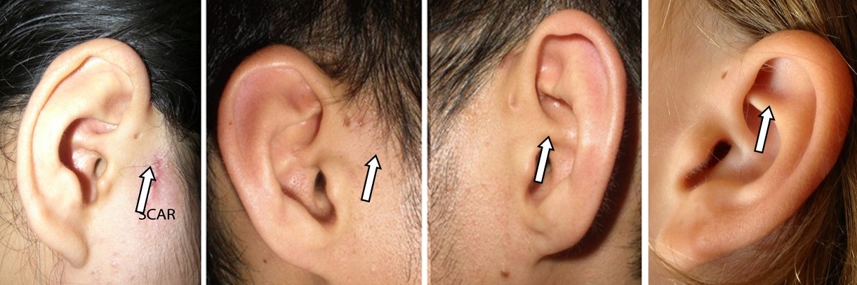 |
| Figure 2: Preauricular Sinus |
Patients with preauricular sinuses may present with facial cellulitis or ulcerations located anterior to the ear.
Clinical presentation
- Pain
- Discharge
- Swelling
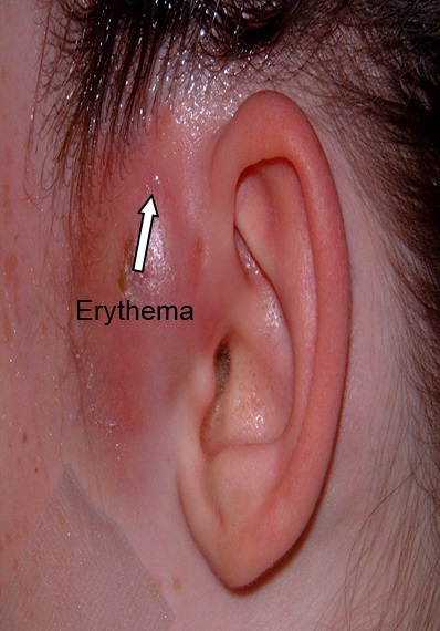
- Erythema /inflammation/ red colour skin
- Ulcer
 |
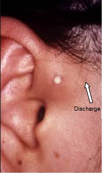 |
| Figure 3: Preauricular abscess | Figure 4: Discharge |
Some cases of preauricular sinuses are associated with other congenital facial deformity syndromes, like Treacher Collins syndrome, branchio-oto-renal syndrome.
Complications
Untreated preauricular sinus itself does not directly lead to any life-threatening conditions.
Complication associated with it includes:
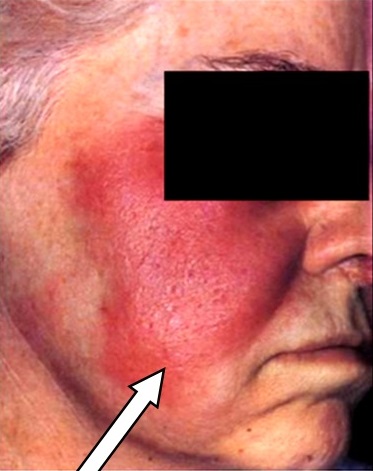
- Facial swelling with red painful skin (facial cellulitis)
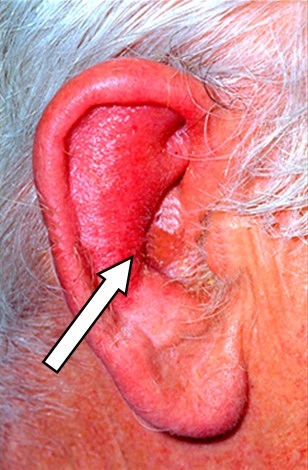
- Painful swelling with redness of the ear (Perichondritis)
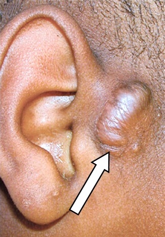
- Abnormal Scar (keloid) – continuous excessive scar tissue formation (hyperlink: Ear Keloid)
- Ear discomfort
 |
 |
 |
||
| Figure 5: Facial Cellulitis | Figure 6: Perichondritis | Figure 7: Keloid |
Treatment
Most preauricular sinuses are asymptomatic and best left untreated. However, if the preauricular sinus becomes repeatedly infected, surgical removal of the sinus is recommended.
Once a patient acquires infection of the sinus, he or she must receive systemic antibiotics. If an abscess is present, it must be incised and drained. The exudates/pus should be sent for bacteria culture to ensure proper antibiotic prescription
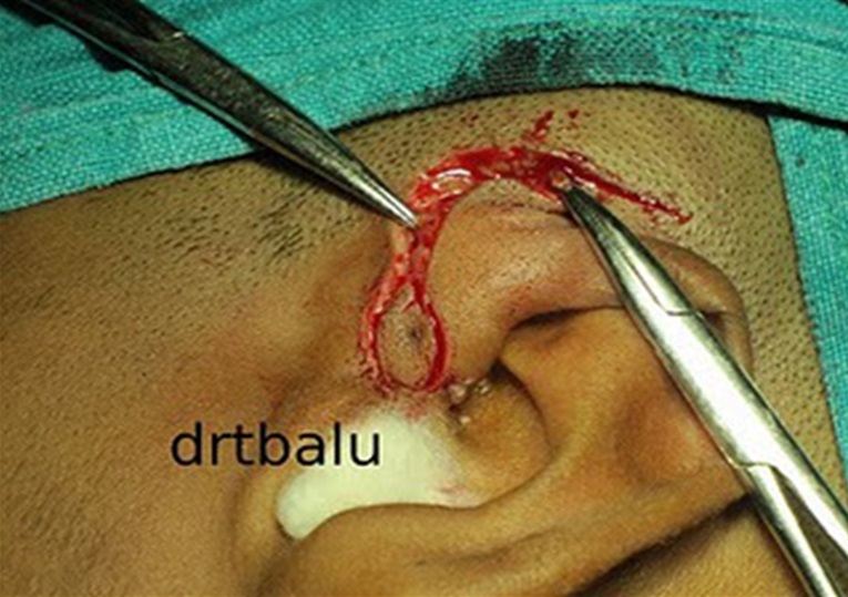 |
| Figure 8: Operation to remove preauricular sinus (Source: https://otolaryngologynews.files.wordpress.com/2011/09/comet.jpg?w=320&h=240) |
Watch Video “Preauricular Sinus Excision”
Prognosis
Preauricular sinuses generally have a good prognosis and outcome.
References
- Amer I, Falzon A, Choudhury N, Ghufoor K. Branchiootic syndrome–a clinical case report and review of the literature. J Pediatr Surg. 2012 Aug. 47:1604-6.
- Diseases of Ear, Nose and Throat, 4th edition (2007), PL Dingra, Elsevier
- Choi SJ, Choung YH, Park K, Bae J, Park HY. The variant type of preauricular sinus: postauricular sinus. Laryngoscope. 2007 Oct. 117(10):1798-802.
- I P Tang, MD, S Shashinder,S Kuljit, K G Gopala : Outcome of Patients Presenting with Preauricular Sinus in Tertiary Centre – A Five year Experience. Med J Malaysia. 2007 March. Vol 62.
- Scheinfeld NS, Silverberg NB, Weinberg JM, Nozad V. The preauricular sinus: a review of its clinical presentation, treatment, and associations. Pediatr Dermatol. 2004 May-Jun. 21(3):191-6.
| Last Reviewed | : | 1 December 2015 |
| Writer | : | Dr. Eshamsol Kamar b. Omar |
| Accreditor | : | Dr. Faridah bt. Hassan |


